Haustorial Initiation in Orobanchaceae
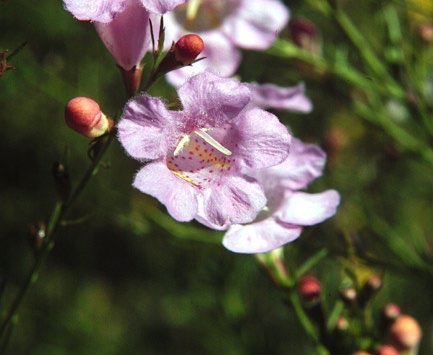
- Agalinis purpurea. This
species served as a model organism for studies of haustorial initiation
in the lab of Dr. James Riopel, University of Virginia in the 1980s and
beyond. Photo by Lytton J. Musselman.
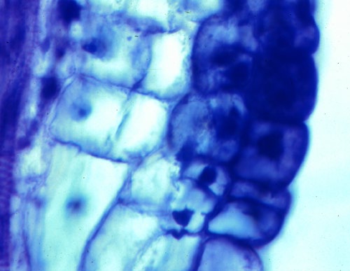
- Agalinis purpurea.
Root, 18 hours after being induced to form a haustorium by xenognosins.
The epidermal cells become densely cytoplasmic and cell divisions begin.
Photo by J. Riopel.
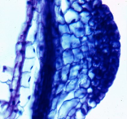
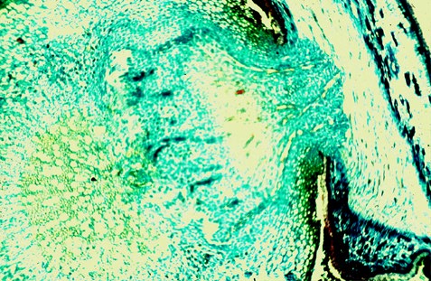
- Agalinis purpurea.
Root, 3 days after being induced to form a haustorium by
xenognosins. Swelling begins below the densely staining epidermal cells.
Photo by J. Riopel.
- Agalinis purpurea.
Root, 6 days after being induced to form a haustorium by
xenognosins. A large swelling is apparent as well as haustorial hairs.
Photo by J. Riopel.
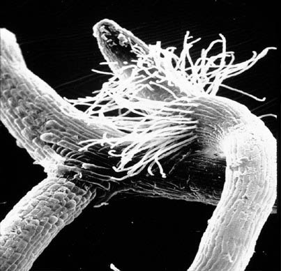
- Agalinis purpurea.
Scanning electron microscope (SEM) picture of haustorial root attaching
to a host root. Photo by Wm. Vance Baird.
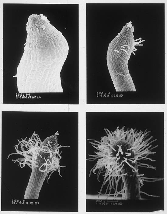
- Agalinis purpurea. SEM
pictures showing series of developmental stages of haustoria. Photo by
Wm. Vance Baird.
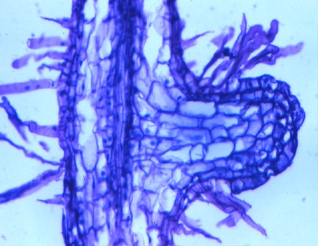
- Aureolaria flava -
mature haustorium in section. Host root is to the right. Photo by L. J.
Musselman.
A related member of Orobanchaceae, Triphysaria versicolor, is
being used as a model organism in John Yoder's lab, University of
California, Davis. Visit his web page HERE
called "Listening to Plants Communicate" to see how the intricate process
of germination and host recognition has been dissected in Tryphysaria.

Last updated: 03-May-18 / dln